Search results for "stains dyes"

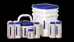
Epredia™ Shandon™ Tissue-Marking Dyes, Tissue marking dye kit
Permanently mark the margins of excised surgical specimens for exact orientation with Epredia™ Shandon™ Tissue-Marking Dyes.

Epredia™ Mark-It™ Tissue Marking Dyes
For a variety of tissue identification purposes use Epredia™ Mark-It™ Tissue Marking Dyes.

Invitrogen™ CellMask™ Plasma Membrane Stains

Invitrogen CellMask™ plasma membrane stains enable fast and uniform labeling of the plasma membrane in almost all cell types. These stains work rapidly and can be detected using standard microscope filter sets with any imaging instrument.

Invitrogen™ DAPI and Hoechst Nucleic Acid Stains
DAPI and Hoechst are classic popular nuclear counterstains for use in all fluorescent cell and tissue techniques. The blue fluorescence is vivid contrast to green, yellow, orange, red, far red and NIR fluorescent probes and labels.
BD Difco™ Stains, Dyes, and Indicators
For Gram stain and acid-fast staining procedures
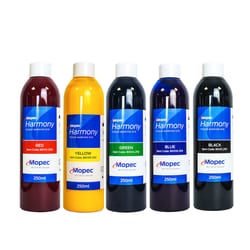
Mopec™ Harmony Tissue Marking Dye Kit
Mopec™’s range of Harmony tissue marking dyes are used to permanently mark surgical tissue margins in Histological specimens. Marking dyes are available in 9 colors, with a standardized viscosity over the range.

Thermo Scientific™ Pierce™ Coomassie Brilliant Blue Dyes
Thermo Scientific Pierce Coomassie Brilliant Blue dyes are composed of one of the most common forms of coomassie dye, which is a key component of various colorimetric protein gel stains.
Molecular Probes™ HCS NuclearMask™ Stains
Easily identify cell nuclei and other features of interest during high-content screening (HCS) assays with the versatile HCS NuclearMask Blue, Red, and Deep Red stains, which can be used with live and fixed cells and in multiparametric analyses.

Mopec™ Harmony Tissue Marking Dye Kit with Stand
Mopec™’s range of Harmony tissue marking dyes are used to permanently mark surgical tissue margins in Histological specimens. Marking dyes are available in 9 colors, with a standardized viscosity over the range.

Invitrogen™ Dimeric Cyanine Nucleic Acid Stains
Dimeric Cyanine Nucleic Acid stains are eight spectrally distinct dyes for ultrasensitive fluorescence detection of nucleic acid in imaging and flow cytometry. They are cell-impermeant and have bright fluorescence signals, low background, and strong binding affinity with nucleic acid.

Invitrogen™ SYTO™ Red Fluorescent Nucleic Acid Stain Sampler Kit - SYTO™ dyes 17 and 59-64 - 50 μL each
For use with fluorescence microscopy and flow cytometry
Invitrogen™ SYTOX™ Green Nucleic Acid Stain - 5 mM Solution in DMSO
Useful indicator of dead cells within a population

Invitrogen™ SYBR™ Safe DNA Gel Stain

Highly sensitive stain for visualization of DNA in agarose or acrylamide gels

Thermo Scientific Chemicals Auramine O, 80% dye content
CAS: 2465-27-2 Molecular Formula: C17H22ClN3 Molecular Weight (g/mol): 303.834 MDL Number: MFCD00012484 InChI Key: KSCQDDRPFHTIRL-UHFFFAOYSA-N Synonym: C.I. 41000 PubChem CID: 17170 ChEBI: CHEBI:51876 IUPAC Name: 4-[4-(dimethylamino)benzenecarboximidoyl]-N,N-dimethylaniline;hydrochloride SMILES: CN(C)C1=CC=C(C=C1)C(=N)C2=CC=C(C=C2)N(C)C.Cl

Invitrogen™ CellTracker™ CM-DiI Dye
A red fluorescent dye well suited for monitoring cell movement or location